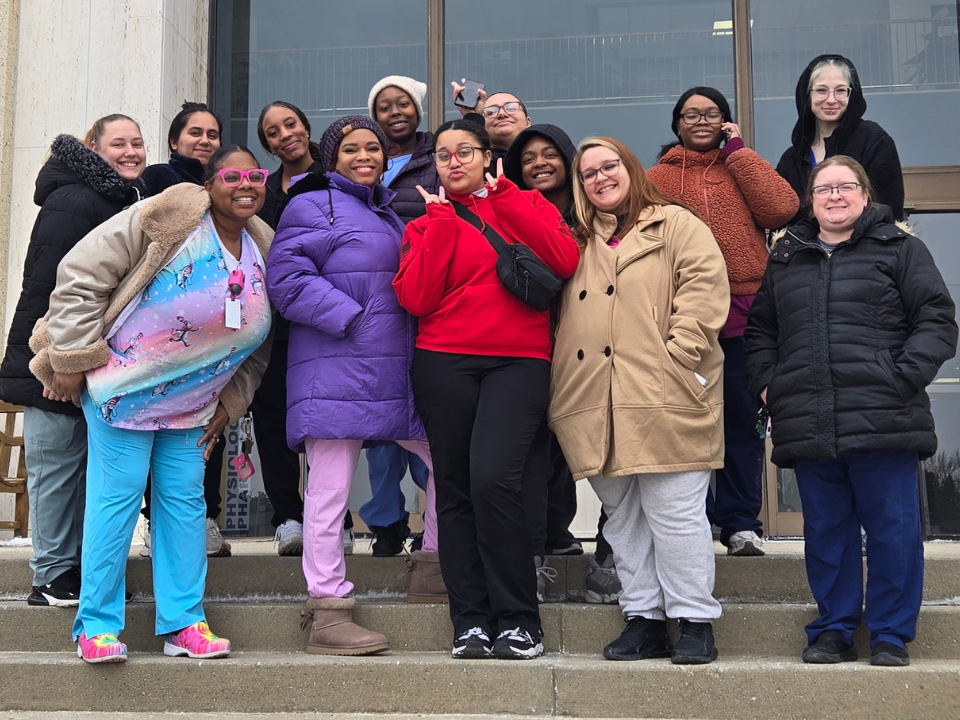
Students and staff from Ross College’s Sylvania campus recently had the unique opportunity to step outside the classroom and into a real clinical learning environment during a visit to the Cadaver Lab and Plastination Museum at the University of Toledo Medical Center.
For Medical Assistant and Dental Assistant students, anatomy and physiology are foundational components of their education. Understanding the human body is essential for assisting providers, supporting patient procedures, and delivering quality care. While textbooks and lectures provide the groundwork, seeing real human anatomy firsthand offers a level of clarity and perspective that cannot be replicated in a traditional classroom setting.
During the visit, students were able to observe and respectfully touch and hold human cadavers, fully dissected organs, and plastinated specimens. The experience allowed them to examine the size, structure, and complexity of various body systems in a way that deepened their understanding and strengthened their confidence.
The Plastination Museum serves as a dynamic study and teaching space designed to support healthcare education. Plastination is a sophisticated, multi step preservation technique invented in 1977 by Dr. Gunther von Hagens. The process replaces water and lipids in biological tissues with curable polymers such as silicone or epoxy, creating dry, durable, and odorless anatomical specimens. These preserved structures maintain remarkable detail, allowing students to clearly view muscles, nerves, blood vessels, and organs.
The museum’s anatomy collection includes prosections arranged by region of the body, giving students a comprehensive and organized way to study human structure. Areas covered include the cranial cavity and brain, head and neck, thorax and thoracic viscera, abdominal cavity and abdominal viscera, spinal cord and vertebral column, as well as the upper and lower limbs. In addition to normal anatomical structures, the collection also features pathological specimens that support the study of disease processes. These specimens are arranged to correspond with the regional displays, helping students better understand how illness and injury affect specific areas of the body.
Stephanie Versellie, Student Success Advisor at the Sylvania campus, shared how meaningful the experience was for students.
“This visit truly brings anatomy to life,” Versellie said. “There is a noticeable difference when students move from studying diagrams to seeing real anatomical structures. It deepens their comprehension and reinforces the level of responsibility they will carry as healthcare professionals.”
Faculty members noted that students were highly engaged throughout the visit, asking questions and making strong connections to concepts covered in class. Seeing the intricate design of the human body firsthand helps solidify knowledge that will be applied in clinical settings.
Opportunities like this help bridge the gap between classroom learning and real clinical environments. By seeing anatomy firsthand, students gain a clearer understanding of the structures they will encounter in patient care settings and strengthen the foundation they are building in their programs.
For the Sylvania campus students, the visit was not just a day away from class. It was an experience that reinforced their purpose, sharpened their skills, and reminded them why a strong understanding of anatomy is so essential in healthcare.